- Noticias / Rapid and cost‐effective process based on insect larvae for scale‐up production of SARS‐COV‐2 spike protein for serological COVID‐19 testing
Rapid and cost‐effective process based on insect larvae for scale‐up production of SARS‐COV‐2 spike protein for serological COVID‐19 testing
Compartir en
redes sociales
Ignacio Smith | Gregorio Juan Mc Callum | Adriana Victoria Sabljic | Juan Ignacio Marfia | Silvina Sonia Bombicino | Aldana Trabucchi | Ruben Francisco Iacono | Joaquín Manuel Birenbaum | Susana Claudia Vazquez| Juan Mauricio Minoia | Osvaldo Cascone | María Gabriela López | Oscar Taboga| Alexandra Marisa Targovnik | Federico Javier Wolman1| Matías Fingermann | Leonardo Gabriel Alonso | Silvina Noemí Valdez | María Victoria Miranda
ABSTRACT
Serology testing for COVID‐19 is important in evaluating active immune response against SARS‐CoV‐2, studying the antibody kinetics, and monitoring reinfections with genetic variants and new virus strains, in particular, the duration of antibodies in virus‐exposed individuals and vaccine‐mediated immunity. In this study, re[1]combinant S protein of SARS‐CoV‐2 was expressed in Rachiplusia nu, an important agronomic plague. One gram of insect larvae produces an amount of S protein sufficient for 150 determinations in the ELISA method herein developed. We es[1]tablished a rapid production process for SARS‐CoV‐2 S protein that showed im[1]munoreactivity for anti‐SARS‐CoV‐2 antibodies and was used as a single antigen for developing the ELISA method with high sensitivity (96.2%) and specificity (98.8%). Our findings provide an efficient and cost‐effective platform for large‐scale S protein production, and the scale‐up is linear, thus avoiding the use of complex equipment like bioreactors.
INTRODUCTION
The outbreak of a new virus, SARS‐CoV‐2, in December 2019 has had a serious impact on human health (Zhu et al., 2020). The severe acute respiratory syndrome related to this virus, officially designated COVID‐19, has rapidly spread all over the world, progressing into a pandemic. This situation has urgently impelled many companies and public research institutes to concentrate their efforts on the search for effective vaccines, therapeutics, and diagnostic tests.
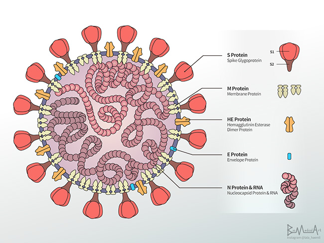
SARS‐CoV‐2 is a single‐stranded RNA‐enveloped virus. The coronavirus spike (S) glycoprotein is surface‐exposed in a large number of copies, and it mediates entry into host cells by interacting with angiotensin‐converting enzyme 2 (ACE2) (Ke et al., 2020). For these reasons, S protein rapidly became the main target of neu[1]tralizing antibodies and the focus of therapeutic and vaccine design (Salvatori et al., 2020). In virions, the S protein exists as a large (over 500 kDa), highly glycosylated homotrimer, each monomer consisting of a globular head, the S1 subunit with its receptor‐binding domain (RBD), and the S2 subunit, containing the protein‐machinery that mediates viral‐cell membrane fusion. The S protein normally exists in a metastable, prefusion conformation, but once the virus interacts with the host cell, an extensive rearrangement occurs, allowing the virus to fuse with the host cell membrane. The spikes are coated with polysaccharide molecules to camouflage themselves, evading sur[1]veillance by the host immune system during entry (Watanabe et al., 2020)
Studies conducted in COVID 19 patients have reported that S and nucleocapsid (NCP) proteins are the main SARS‐CoV‐2 antibody targets (Burbelo et al., 2020). These antibodies are detectable from approximately 6 days after PCR confirmation of infection. It was demonstrated that those antibodies directed against RBD into S show a neutralizing capacity and, hence, can prevent infection (Seydoux et al., 2020; Suthar et al., 2020)
Serological tests for COVID‐19 are based on the detection of multiple targets of the virus, including S, RBD, NCP, and nonstructural proteins, and are extensively used to identify whether people have been exposed to SARS‐CoV‐2 by looking at their im[1]mune response (Ghaffari et al., 2020). During the pandemic, many efforts have been directed towards detecting, tracking, and better understanding human humoral responses to SARS‐CoV‐2 infection. It is crucial to develop robust and reliable serological assays to study the antibody kinetics and neutralization efficiency and monitor re[1]infections with genetic variants and new virus strains, in particular, the duration of antibodies in virus‐exposed individuals and vaccine‐ mediated immunity. Currently, the RBD and S proteins are the most reliable antigens for measuring the abundance of neutralizing anti[1]bodies (Galipeau et al., 2020)
Different strategies were described to obtain the S protein by biotechnological methods. Because of the structural complexity and posttranslational modifications of the S protein, major efforts were directed to mammalian cell culture as a suitable productive system (Stuible et al., 2021). As already described, the S protein is a large homotrimer with 22 N‐linked glycosylation sites per monomer (Watanabe et al., 2020). As a consequence of its structural com[1]plexity, it is not surprising systems like Chinese hamster ovary and human embryonic kidney render low yields in mammalian (Esposito et al., 2020; Walls et al., 2020). Moreover, the great volume of culture media needed for S expression in mammalian cultures at a high scale is too expensive, especially to obtain this antigen for serological assays.
We previously exhaustively studied the use of insect larvae such as Rachiplusia nu, an important agronomic plague in America, as a platform to produce different proteins in a short time and at a low cost (Targovnik et al., 2016). Also, we previously identified and de[1]scribed the chromatographic behavior of the main contaminant proteins present in the host to facilitate the downstream processing of any recombinant protein produced in this system (Mc Callum et al., 2019)
Here, we established a rapid and cost‐effective process for the expression and purification of a high‐quality trimeric version of SARS‐CoV‐2 S protein by using the baculovirus‐insect larvae system. This novel recombinant protein was used for developing a new serological ELISA test for COVID‐19, showing high sensitivity and specificity with low operational complexity and cost.
MATERIALS AND METHODS
Recombinant virus construction
We based our studies on a previously described version of SARS‐ CoV‐2 S protein sequence, stabilized in its prefusion conformation (Wrapp et al., 2020). Briefly, the expressed protein was expected to include the ectodomain (residues 19‐1207) of the SARS‐CoV‐2 Wuhan‐Hu‐1 S protein (GenBank: QHD43416.1) without native signal peptide, where the furin cleavage site (residues 682–685, PRRA) was removed (GSAS mutations) and residues at positions 986 (K) and 987 (V) were mutated to proline; additionally, a C‐terminal T4 fibritin trimerization domain, a TEV protease cleavage site, and a histidine tag were included (Figure 1). For secretion, S protein was expressed under gp64 baculoviral signal peptide. The DNA sequence was codon‐optimized for baculovirus expression and chemically synthesized (Genscript). The S sequence and the enhanced green fluorescent protein (EGFP) cDNA were cloned into the pFastBacDual vector (Thermo Fisher Scientific) under the polyhedrin (polh) and p10 promoter, respectively, for expression in the baculovirus system. For this purpose, the EGFP (GenBank Accession no. NC_013179.1) was cloned into SmaI and Nco1 sites of the pFastBacDual vector (Targovnik et al., 2019)]. Then, the S cassette was subcloned into the pFastBacDual Vector into BamH1 and HindIII sites to generate the pFBD‐p10‐EGFP‐polh‐S construction.
The recombinant baculoviruses were obtained using the Bac to Bac® baculovirus expression system (Thermo Fisher Scientific), following the manufacturer's instructions. The pFBD‐p10‐EGFP‐polh‐S vector was transformed into a chemically competent Escherichia coli DH10Bac™ strain (Thermo Fisher Scientific) by heat shock to generate the recombinant bacmid by transposition. Then, the bacmids were purified and used to transfect 1 million Sf9 cells using Cellfectin II Reagent (Thermo Fisher Scientific). After 4‐day incubation at 27°C, the cell culture supernatant was collected and centrifuged at 500g for 10 min. The transfection efficiency was determined by measuring EGFP expression by fluorescence under UV light. The recombinant Autographa californica nuclear polyhedrosis virus (AcMNPV) polyhedrin‐minus virus containing EGFP under the control of the p10 promoter and the S sequence under the control of the polyhedrin promoter was named rAcMNPV‐S. Then, a round of amplification was performed in Sf9 cells seeded in T‐25 flasks at 27°C, at a low multiplicity of infection (MOI) of 0.02. The Sf900 II insect cell culture medium and the antibiotic and antimycotic solutions were from InvitrogenTM (Gaithersburg) and the fetal bovine serum was from Nutrientes Naturales S.A. (Buenos Aires). The amplified rAcMNPV–S was titrated by plaque assay (O'Reilly et al., 1994). This high‐titer rAcMNPV–S was the viral stock used for protein production in insect larvae.
Expression of S protein in insect larvae
nu larvae were from AgIdea (Pergamino). They were reared in trays at 23–25°C in a 70% humidified chamber, with a 16:8 light:dark photoperiod, and fed a high wheat‐germ diet until they reached their fifth instar (20 days of age) prepared by AgIdea (Pergamino). For all the experiments, batches of 500 fifth‐instar larvae were injected with 50 µl of the recombinant baculovirus stock (diluted to 1 × 10⁷ PFU ml−1) near the third prolegs, as shown in Figure 2. To char[1]acterize and quantify the recombinant protein produced, larvae that were alive and fluorescent under UV light were harvested at Day 4 postinfection and frozen immediately at −80°C until they were processed for analysis. Larvae infected with a nonrelated re[1]combinant baculovirus were included as the control.
Recombinant S protein purification by immobilized metal‐ion affinity chromatography (IMAC)
In a typical process, 75 g of infected larvae were homogenized directly with 200 ml of equilibration buffer containing 10 mg glutathione crystals, 50 mM arginine, and 4 mM PMSF and 1/200 (vol/ vol) protease inhibitor cocktail (Sigma‐Aldrich) using a Bag Mixer 400 homogenizer (Interscience). Then, the larval extract was centrifuged at 10,000g for 30 min at 4°C and the pellet was discarded. The supernatant was filtered through Whatman paper using a filter holder with a receptor (Nalgene) to remove the lipid fraction remaining at the top. The filtered sample was centrifuged for a second time in the same conditions, and the supernatant was filtered through 3 µm (Sartopore o SartoBran capsules). Then, two experiments were conducted. In the first experiment, the clarified homogenate was loaded into the HisTrap FF 5 ml column (Cytiva) previously equilibrated with 10CV (column volume) of 20 mM phosphate buffer, pH 7.4, 20 mM imidazole, 300 mM NaCl, and 50 mM arginine, and washed with 10CV of the same buffer. The recombinant protein was eluted with a buffer containing 500 mM imidazole, 100 mM arginine, and 10% glycerol. In the second experiment, the same chromatography was done but the matrix was equilibrated with the same buffer containing 20 mM imidazole, a second wash was done with 80 mM imidazole, and then the S protein was eluted with 500 mM imidazole. After that, an optimized protocol consisted in equilibrating the chromatographic matrix directly with the same buffer containing 80 mM imidazole, and after washing, the S protein was eluted by adding 500 mM imidazole. In all cases, the linear flow rate was 0.4 cm/min, and all fractions were collected and subjected to sodium dodecyl sulfate–polyacrylamide gel electrophoresis (SDS‐PAGE) and western blot analysis
Determination of protein concentration Total protein concentration
Was determined by the Bradford microassay protocol (Bradford, 1976) using the Quick Start™ Bradford reagent (BioRad). The samples used were crude larval extract and the purified S fraction. Additionally, the concentration of purified S protein was determined in an SDS‐PAGE image with a diluted series of control bovine serum albumin.
SDS‐PAGE and western blot analysis
Larval extracts and purification fractions were resolved by SDS‐ PAGE (10% polyacrylamide gels). Before loading the samples into the wells, they were heated for 5 min at 100°C in sample buffer (125 mM Tris–HCl, pH 6.8, 4% [wt/vol] SDS, 20% [wt/vol] glycerol, 0.01% [wt/ vol] bromophenol blue, and 10% [vol/vol] 2‐mercaptoethanol). One of the lanes was reserved for the protein marker to determine the MW of the protein bands. The resulting gels were either stained with Coomassie Blue R‐250 or transferred onto nitrocellulose membranes (Cytiva). Membranes were then incubated overnight at 4°C in blocking solution (phosphate‐buffered saline [PBS]‐3% skim milk [PBS‐M]). After a 10‐min wash with PBS containing 0.05% vol/vol Tween 20 (PBS‐T), the membrane was incubated for 2 h with mouse anti‐His antibody (BD Biosciences) 1/2500 in 0.05% PBS‐T‐1% skim milk, and then washed three times with PBS‐T. Polyclonal rabbit anti‐ mouse immunoglobulin conjugated with HRP (1/30000 in 0.05% PBS‐T‐1% skim milk) was used as the secondary antibody. Development was carried out with 3,3'‐diaminobenzidine (Sigma‐Aldrich) staining or, alternatively, with an enhanced chemiluminescent substrate (ECL) and high‐performance chemiluminescence films (CL‐X Posure™; Thermo Fisher Scientific)
Characterization of the recombinant S protein expressed in R. nu
Size exclusion chromatography
The oligomerization state of the recombinant S protein was evaluated by high‐resolution size exclusion chromatography (SEC). Elution fractions from IMAC containing purified S protein were collected and concentrated, and the buffer was exchanged and loaded on a Superdex 200 increase 10/300 (Cytiva) equilibrated in 100 mM sodium phosphate buffer, 150 mM NaCl, pH 7.4. All fractions were collected, and the S protein was revealed by WB using an anti‐6x His‐tag antibody, as described previously. The calculated molecular weight (MW) of the expressed protein, without considering the glycan moieties, was 138914 Da.
Sera collection
Healthy control individuals
Control serum/plasma (n = 83) was obtained from samples collected from healthy individuals before the outbreak of SARS‐CoV‐2. The sample collection was approved by the Ethics Committee of José de San Martín Clinical Hospital, University of Buenos Aires (UBA), Buenos Aires, Argentina. Sera were stored at −20°C until assayed
COVID‐19 patients
Serum/plasma samples were collected from a total of 98 COVID‐19 cases confirmed to be infected with SARS‐CoV‐2 by real‐time reverse transcription‐polymerase chain reaction (rRT‐PCR) on samples from the respiratory tract. These samples were provided by the Biobank of Infectious Diseases (BBEI) of the Institute for Biomedical Research on Retroviruses and AIDS (INBIRS). Seventy‐eight of these samples were IgG positive and 20 were IgG negative for SARS‐CoV‐2 by COVIDAR IgG ELISA test (Laboratorio Lemos S.R.L.). Sample collection and protocols were approved by the Ethics Committee of BBEI‐INBIRS and the Ethics Committee in Clinical Research of the School of Pharmacy and Biochemistry, UBA. All subjects were informed about the purpose of the study, and they signed consent for study participation. Sera were stored at −20°C until assayed
Spike application in an immunoassay for antibodies for SARS‐CoV‐2 assessment
Reagents
PBS was used as the microplate coating buffer. PBS‐M and PBS‐T were used as blocking solution and washing buffer, respectively. Sample or reagent dilutions were prepared in 3% wt/vol skim milk, 0.05% vol/vol Tween 20 in PBS (PBS‐MT). Streptavidin–Horseradish peroxidase (HRP) and rabbit anti‐human IgG‐biotin were purchased from Jackson ImmunoResearch Laboratories, Inc. The 3,3',5,5'‐tetramethyl‐benzidine/ 4 | SMITH ET AL. H2O2 (Single Component TMB Peroxidase EIA Substrate Kit; BioRad) was employed as the chromogenic substrate. Except when otherwise indicated, incubations were performed at RT, washing steps were per[1]formed with PBS‐T, and 50 μl per well were added in each incubation step
ELISA with colorimetric detection
Polystyrene microplates (Maxisorp; NUNC) were coated overnight at 4°C with 0.1 μg purified Spike per well, in coating buffer, and washed five times with PBS. Blocking solution (200 μl/well) was added, and plates were incubated for 1 h. After washing six times, duplicate serum samples diluted 1/100 (100 μl/well) were added and incubated for 1 h. Microplates were washed six times and incubated for 30 min at 37°C with anti‐human IgG‐biotin (diluted 1/180000). Plates were washed six times, and bound antibodies were detected with Streptavidin‐HRP (diluted 1/2000, 30 min at 37°C). After washing (five times plus one final wash with PBS), the chromogenic substrate was added and plates were incubated for 15 min in the dark. The color reaction was stopped with 2 M H2SO4. The oxidized substrate was measured at 450 nm with an ELISA plate reader MultiskanFC (Thermo Scientific Labsystems). The blank control was made by replacing serum samples with PBS‐MT. Results were calculated as specific absorbance (A = the mean of each sample minus the mean of the blank control) and expressed as stan[1]dard deviation scores: SDs = (A‐Ac)/SDc, where Ac is the mean specific absorbance of healthy control sera and SDc its standard deviation. The cut‐off value of the assay was set at SDs = 3.0
Statistical analysis
The normal distribution of data was analyzed by the D'Agostino and Pearson omnibus normality test. To remove outliers from normally distributed healthy control individuals, the Rout test was performed. The selection of optimal cut‐off values was based on curves constructed by plotting the calculated specificity and sensitivity versus the correspond[1]ing cut‐off values. Statistical significance was evaluated using either parametric tests: paired‐samples Student t test and unpaired‐samples Student t test with Welch correction, or nonparametric tests: Wilcoxon matched‐pairs signed‐rank test or Mann–Whitney U test for unpaired data, when applicable. Calculations were performed using GraphPad Prism version 6.01 for Windows (GraphPad Software; www.graphpad. com). A p value < 0.05 was considered statistically significant.
RESULT AND DISCUSSION
Expression and purification of the recombinant spike in insect larvae
A low‐cost alternative to cell culture‐based protein production is the use of live insect larvae as “mini bioreactors.” To obtain the recombinant version of the S protein, we cloned the expression cassette into the pFastBacDualGFP vector under the control of the strong baculovirus polyhedrin promoter. The expression cassette included gp64 signal peptide, which targets the recombinant protein to the secretion pathway. After transfection and amplification in Sf9 insect cells, a recombinant baculovirus seed‐stock for expression in larvae was obtained. Insect larvae support many of the posttranslational modifications that enable proteins to achieve their biologically functional native conformation (Loustau et al., 2008)
nu larvae were infected by injection of the baculovirus by intrahemocele injection with approximately 5 × 10⁵ pfu of viral stock (Figure 2a,b). From our experience, this dose was the best option to achieve a high level of S protein expression and no larval mortality. The larvae infected with the recombinant baculovirus expressed, in addition to the proteins of interest, the EGFP protein, which allowed us to determine the optimum day of harvest by observation under UV light. At 4 day postinfection (dpi), the fluorescence was maximal, and then the viability of the larvae decreased significantly. Therefore, the larvae were harvested at 4 dpi under a UV lamp using fluorescence as an indicator of infection. We proceeded to obtain the clarified homogenate from the larvae and purify the recombinant proteins expressed in insect larvae R. nu by IMAC. The gp64 signal peptide was effective to target S protein to the secretory pathway. The recombinant S protein was secreted to hemolymph and this localization made it easier to extract it from the larvae.
SDS‐PAGE revealed that most of the proteins of the crude extract were removed in the passthrough fraction during purification by IMAC when the sample was loaded without imidazole. However, when the bound material, containing the protein of interest, was desorbed after a single 500 mM imidazole step, an important contaminant of hemolymph (hexamerin, 76 kDa approx.) was still present in the elution fraction. After some rounds of optimization, we concluded that when the extracts were run directly on the chromatographic matrix previously equilibrated with 20 mM imidazole, most contaminants, including hexamerin, eluted in the passthrough or washing fractions, while the recombinant S protein remained bound to the matrix. Nevertheless, in this condition, another contaminant with a molecular weight similar to that of the S protein was present and eluted with 80 mM imidazole (Figure 3). For these reasons, we assessed a new protocol, which directly equilibrated the matrix with the same buffer containing 80 mM imidazole. As judged by reducing SDS‐PAGE and WB, the estimated molecular weight of the recombinant S protein was 150 kDa (monomer), indicating that it was correctly glycosylated and did not suffer protease degradation (Figure 3). Other authors have expressed the ectodomain of S protein with the native peptide sequence in hemolymph of Bombyx mori larvae and reported that recombinant protein was cleaved probably by a host furin‐protease. In the same work, the authors resolved it with a version of S protein modified in furin protease‐target site (Fujita et al., 2020). In the present work, we decided to synthesize a version of S protein where the furin cleavage site (residues 682–685, PRRA) was removed because furin protease activity was only described in Spodoptera frugiperda, an R. nu related species (Westenberg et al., 2002)
The amount of recombinant S protein was 15 ug/g of larvae at 4 dpi on our platform based on R. nu. The process based on B. mori previously reported by other authors (Fujita et al., 2020) can be compared with ours as follows: in both cases, the optimal day of S protein harvest was 4 dpi after larval infection; however, in B. mori, it was necessary to extract the whole hemolymph of each larva for purification while, in our case, a complete extract was done with all infected larvae, thus simplifying the biotechnological process. In B. mori, the estimated value of the purified S protein from 10 ml hemolymph (35 larvae) was 100 µg, and the same yield was obtained with 45 R. nu larvae in the process herein described.
Protein quality assessment
We assessed whether insect larvae‐produced SARS CoV‐2 S protein was suitable for our intended use. The trimetric nature of the S protein, which is typical of Type I viral fusion proteins, is a critical quality attribute of recombinant S proteins in different expression systems (Esposito et al., 2020). We evaluated the oligomeric state of the IMAC‐purified S protein from R. nu larvae by SEC in a Superdex 200 column. Figure 4a shows a typical SEC chromatogram of the larvae‐expressed SARS CoV‐2 S protein eluting as a broad peak with an elution volume close to 10 ml, compatible with a 450–500 kDa protein, in good accordance with the expected trimeric nature of the purified protein. The S monomer is a protein with an apparent molecular weight of ∼150 kDa bearing many glycan moieties, which affects its hydrodynamic behavior (the MW calculated from the amino acid sequence is 138 kDa). The presence of S protein in these fractions was confirmed by WB, using an anti‐6x His tag‐specific antibody (Figure 4b). Notably, two peaks, denoted as Peak #1 and #2, were also observed. However, these peaks were not recognized by the anti‐6x His tag antibody in the WB revealed with ECL (Figure 4b), indicating that they might correspond to nonrelated impurities or to a dissociated and fragmented S protein with its carboxyl‐terminal cleaved. It should be noted that some proteolysis can occur during concentration and buffer exchange of the sample performed before injecting on an SEC column
SPIKE application in an immunoassay for antibodies for SARS‐CoV‐2 assessment
The immunochemical behavior of the recombinant version of the S protein expressed in insect larvae R. nu was evaluated during the following use as an immobilized antigen in the development of an indirect ELISA aimed at detecting the presence of anti‐S specific IgG antibodies in human serum/plasma samples. After an initial round of optimization (antigen concentration and binding conditions, primary and secondary antibodies dilution, washing steps), a panel of 83 human control serum/plasma and 98 COVID‐19 patients' serum/ plasma were analyzed. To calculate the coefficient variation, a positive serum from a COVID‐19 patient was employed. The intra‐assay coefficient variation was 3.21% (n = 2) and the inter‐assay coefficient variation was 20.11% (n = 4). The test performance was optimized in terms of sensitivity and specificity by evaluating the effect of different cut‐off values (in SDs) on receiver operating characteristic curves (ROC) (Figure 5). The area under the ROC curve (AUC) was 0.893, indicating that the method had high accuracy to distinguish between samples from the two groups under study (Carter et al., 2016). When a cut‐off value of 3.0 SDs was established, 75 out of 78 COVID‐19 samples that tested positive by COVIDAR IgG ELISA Test, were also positive by our developed ELISA (sensitivity: 96.2%). The specificity, calculated as 100% minus the percentage of true negative samples (normal human sera, n = 83) detected as positive, was 98.8%. The median SDs range of true negative samples was −0.22 (range: −1.78 to 3.21) and the median SDs range of true positive samples was 7.32 (range: 0.00–23.20). Moreover, the concordance between the ELISA developed herein and the COVIDAR IgG ELISA test was 95.96%, with a kappa statistic of 0.879, representing a substantial agreement between the two methods. The results presented in the present work demonstrate that the Spike protein produced in insect larvae is immunoreactive against the sera from COVID‐19 patients and can be used in immunoassays for the detection of anti‐SARS‐Cov‐2 antibodies (Figure 6). The strategy reported herein should be accessible to many laboratories and should allow the easy production of Spike. In turn, the availability of properly folded Spike would encourage researchers to improve current and develop new immunochemical tests for anti‐SARS‐Cov‐2 antibodies detection.
On the other hand, it is known that the cellular immune response to SARS‐CoV‐2 is critical in controlling disease. For this reason, it is important to analyze the magnitude and phenotype of the SARS‐CoV‐2‐ FIGURE 5 Analysis of ELISA performance resulting from the study of 83 sera from normal control individuals and 98 sera from COVID‐19 patients. (a) Sensitivity curve (o) and specificity (×) as a function of the possible cut‐off values. The vertical dashed line indicates the cut‐off value with the optimized sensitivity and specificity parameters (cut‐off = 3.0). (b) ROC curve analysis of ELISA, AUC is included FIGURE 6 Antibodies anti‐SARS‐CoV‐2 results obtained by ELISA in sera from normal control individuals and sera from COVID 19 patients IgG positive and negative by COVIDAR IgG ELISA test. Results are expressed as SDs. The cut‐off value (SDs > 3.0) is indicated by a dotted line and medians for each population are indicated as a full line (***p < 0.0001). The values for the dots found on the cut‐off line are as follow: controls subjects (n = 3): 2.81, 3.21, and 2.86; Patients COVIDAR IgG+ (n = 1): 2.81 and Patients COVIDAR IgG− (n = 1): 3.17 SMITH ET AL. | 7 specific T cell response. For this purpose, different authors have employed peptide pools from a range of viral proteins, including spike, nucleoprotein, and membrane protein (Zuo et al., 2021). Additionally, other authors have evaluated T‐cells proliferation after stimulation with these proteins (Avendaño‐Ortiz et al., 2020). In view of that, S protein produced in insect larvae could be also employed for this purpose.
CONCLUSIONS
In this study, we show that SARS‐CoV‐2 recombinant S protein could be produced inexpensively after infection of R. nu larvae and easily purified later after a single chromatographic step. Noteworthy, the antigenic properties of this large, complex, highly glycosylated protein are retained, as evidenced during its application for developing a serologic ELISA test. One gram of insect larvae produces an amount of S protein sufficient for 150 determinations in the ELISA method. The scale‐up of S protein production is linear in this biotechnological platform, avoiding the use of complex equipment like bioreactors. Thus, it is straightforward to con[1]clude that our approach represents a rapid, easy, and cost‐effective method for the production of recombinant S for diagnostic applications. We expect that our approach will bring new tools for serologic test producers to face the unprecedented demand for these products during the current COVID 19 pandemic